Imaging and Analysis focuses on:
Light Microscopy
RBGE laboratories are equipped with stereo and compound microscopes.
Basic stereo microscopes without cameras are available in all laboratories, for plant and fungal observation. Stereo microscopes with CCD cameras are also available for data capture, from snapshot to Z-stack image processing.
A range of research-grade compound microscopes are available for morphological research and for cytology; these are equipped with dark field, phase contrast and differential interference contrast (DIC). These facilitate 3D vision without requiring staining. Our epifluorescent microscope facilitates observation of autofluorescence and fluorescent dye staining-based methodologies.
Our microscope laboratories are equipped with histological processing facilities for paraffin, resin and frozen sectioning applications, as well as tissue clearing methods that allow observation of the inner structures of plants.
Scanning Electron Microscope
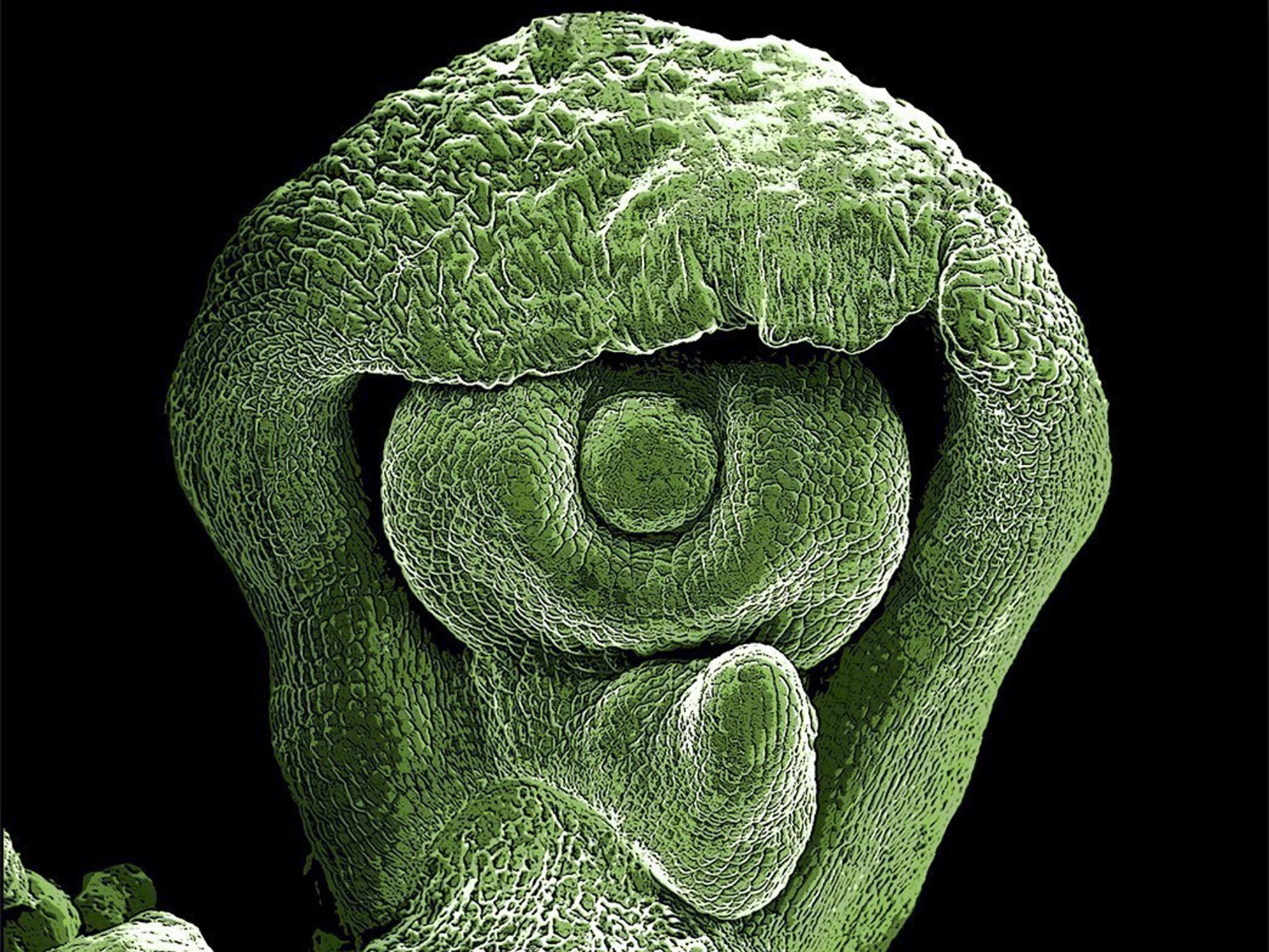
Access to a field emission scanning electron microscope (SEM) provides an opportunity to study plant and fungal surface structures in minute detail, impossible to see with the naked eye or even with compound microscopes. This is useful in the identification and classification of species. For example, pollen characteristics, indumentum, and stomatal features can serve as species’ unique characteristics. The SEM is also intensively used for developmental studies, where ontogenetic series of developmental stages from single cells to young organs can be followed.
SEM images show nature’s art: Dust-like pollen grains come in various shapes, spherical to star-shaped. Leaf surface structures are a mosaic of trichomes and stomata in an abundance of forms and combinations. Former RBGE microscopist Frieda Christie was presented the 2022 Linnean Society Award for her wonderful SEM images, which can be seen in the John Hope Gateway and laboratories.
RBGE has a long history of SEM work, with the first electron microscope installed in 1974. Our fourth SEM was a LEO Supra 55VP which, unlike its predecessors, had a field emission gun producing a beam 1000 × brighter than conventional SEMs. This allowed the observation of very delicate material at lower voltages without loss of detail, and enhanced the depth of field, greatly improves resolution, with up to 900,000 times magnification
Sadly, our LEO Supra 55VP SEM reached the end of its life at RBGE in February 2025. The machine served more than 100 botanists for over 23 years and provided data for numerous publications. We are currently investigating funding and collaboration opportunities to enable access to a SEM.